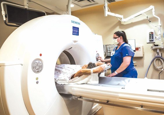
Chest CTs ‘better’ for cancer screening

With lung cancer cases rising in Kenya and globally, citizens have been urged to embrace the use of advanced imaging techniques like chest CT and Positron Emission Tomography (PET) scans for screening.
These techniques can detect the disease early and evaluate how well treatments are working, experts say.
Early detection is crucial in improving survival rates, as it allows for timely intervention and more accurate treatment planning.
“Despite advances in imaging technology, diagnosing lung cancer remains challenging due to the potential for false positives and negatives,” says Dr Solomon Mutua, a clinical oncologist at
Nairobi West Hospital.
“Benign lung nodules can sometimes resemble malignant tumors on a chest CT scan, leading to misdiagnosis.”
He added that “lung cancer can also be hidden within inflammatory changes or scarring in the lungs”.
“That is why use of advanced imaging techniques is recommended to enhance detection and accuracy.”
While a chest X-ray is the quickest, most cost-effective and commonly used initial imaging test for the lungs, it has limitations compared with advanced techniques. A chest X-ray can identify some lung tumors but may miss small, early-stage one and is less effective at assessing cancer spread.
A chest CT scan, on the other hand, provides detailed images by taking multiple pictures of the lungs and chest, and compiling them into a comprehensive view. This imaging allows for the detection of very small nodules and is especially useful for diagnosing lung cancer at its most treatable stage.


