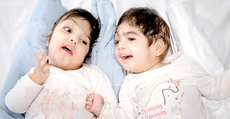
Safa and Marwa Ullah: Successfully separated conjoined twins
By Ann Wairimu, August 1, 20191. They shared a skull and brain tissue
Last month, conjoined twins fused at the head were successfully separated in a London Hospital after a months-long medical endeavour that required more than 50 hours of multiple surgeries and a team of 100 medical professionals.
The two-year-old twins, Safa and Marwa Ullah shared a portion of their skull and brain tissue. Doctors at the Great Ormond Street Hospital used the latest technology, including virtual reality to create exact replicas of the girls’ skulls, and 3D printing to create models to practice on.
In the final operation, doctors built the girls new skulls using their own bones.
2. Kenyan twins shared a spinal cord
Successful separation of two-year-old conjoined twins at Kenyatta National Hospital in November 2016 marked a medical milestone in Kenya.
Kenya became the first country in Sub-Saharan Africa to successfully do such an operation. The procedure took more than 50 medical experts and 23 hours to complete. Favour and Blessing shared a spinal cord, rectum, anus, some muscles, subcutaneous tissues and skin.
The twins had lived together for two years and two months. The surgery was done in three stages — to separate, to close the wounds and to create stomachs for them. They also had to undergo four more reconstructive surgeries after their wounds were healed.
3. Bhutanese twins shared a liver
Last year, Australian doctors successfully separated 15-month-old girls, Nima and Dawa Pelden, who had been joined at the torso and shared a liver.
They had grown facing each other, and could not sit down together. Also, they could only stand at the same time. About 18 specialists in two teams, one for each girl, took part in the procedure at Melbourne’s Royal Children’s Hospital. Doctors successfully separated the twins’ liver.
4. Joined at the lower chest and abdomen
Maria and Teresa were born joined at the lower chest and abdomen, sharing a liver, pancreas and portion of the small intestine.
The girls, from the Dominican Republic, were separated at the Children’s Hospital of Richmond at Virginia Commonwealth University in November 2011.
In several procedures involving six surgeons, the medical team divided the liver, pancreas and other shared organ systems and reconstructed the girls’ abdominal wall.
5. Joined at the liver and chest
At only eight days, twins Lydia and Maya, who were joined at the liver and chest were separated by doctors in Switzerland in early 2016. It took a five-hour operation to separate the girls who were born two months prematurely.
They are believed to be the youngest ever to survive such surgery. The doctors had initial plans to separate the girls after few months.
However, their conditions started deteriorating a week after their birth. One of the twins had high blood pressure and the other had low blood pressure, forcing the doctors to act.
6. Shared a head
Rital and Ritag Gaboura, Sudan-born twin girls were joined at the head. Because Ritag’s heart provided most of the blood for both her and her sister, the girls were given “10 million-to-one” odds of survival.
The surgery took place in four stages in mid-2011; the girls underwent two operations in May and, one month later, tissue expanders, which would help to stretch the babies’ skin over their heads, were inserted.
In August, the final separation occurred and the one-year-old girls showed no adverse reactions.
7. They had one pair of leg
Irish boys Hassan and Hussein Benhaffaf who were joined from the pelvis to the chest, were given only a two per cent chance of survival after their birth in December 2009.
While the twins each had their own heart, the organs shared the same safety “sac,” making the separation all the more dangerous. In addition, the boys, who have only two legs between them, had to have their liver, gut, and bladder separated as well.
But in April 2009 a team of 20 medics, including four surgeons and four anaesthetists working in shifts, operated on the infants for 14 hours. And thanks to replacement limbs, they now walk on their own.
8. Fused at the head
Trishna and Krishna who were born joined at the head, were saved from a Bangladeshi orphanage by their guardian, Moira Kelly.
In 2009, at almost three years of age, the twins underwent a 27-hour operation performed by up to 16 surgeons at the Royal Children’s Hospital in Melbourne, Australia.
While the girls were given just a 25 per cent chance of making it through the surgery alive, they showed signs of health soon after. Ten years later, the twins, who live with their guardians in Melbourne, continue to grow and thrive.
9. Shared liver and diaphragm
Angelica and Angelina Sabuco, Philippine-born twin sisters were joined at the chest and abdomen and shared liver and diaphragm.
On November 1, 2011, the two-year-olds endured a nearly 10-hour surgery by a team of 20 doctors who both separated and reconstructed the area where the girls had been fused.
After the surgery, the girls flourished and within two weeks had normally functioning livers, were off pain medication, and were able to leave the hospital at Stanford University in Palo Alto to begin outpatient therapy.
10. Fused at the lower abdomen
On November 7, 2012, when Amelia and Allison were eight months old, they underwent a marathon surgery. The twins — who were joined at the lower chest and abdomen shared their chest wall, diaphragm, pericardium, and liver.
The seven-hour procedure was tightly orchestrated, with more than 40 physicians, nurses, and other medical staff from general surgery, plastic and reconstructive surgery, cardiac surgery, anaesthesiology, radiology and neonatology participating.
